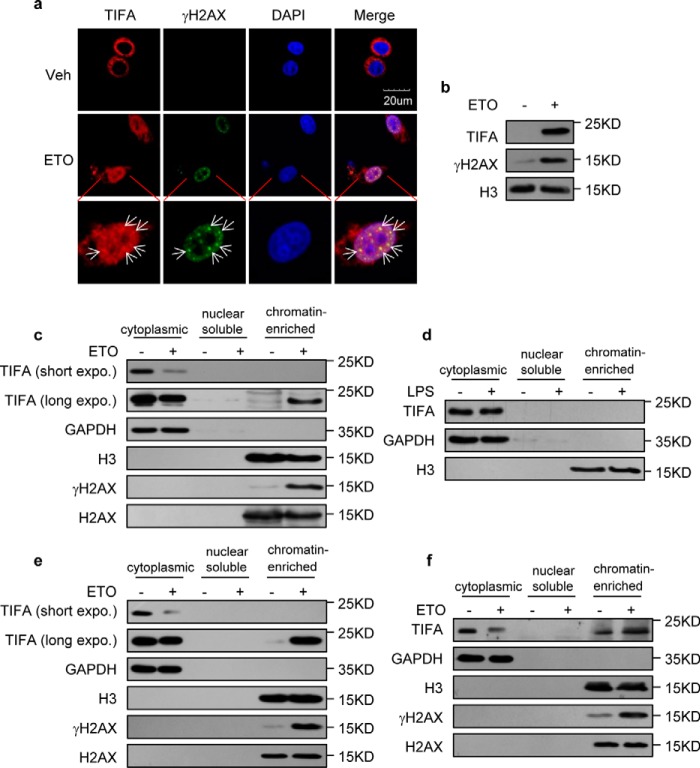
Figure 1.
Enrichment of TIFA on chromatin following DNA damage. a, confocal microscopic examination of TIFA and γH2AX in HeLa cells transfected with FLAG-TIFA were treated with vehicle (Veh) or ETO. 4′,6-Diamidino-2-phenylindole (DAPI) was used to visualize the nucleus. Scale bar represents 20 μm. b, chromatin fractions were isolated from the HeLa cells expressing FLAG-TIFA in the absence or presence of ETO. These fractions were then subjected to Western blotting with the indicated antibodies. c, chromatin fractions were isolated using nuclear lysis buffer containing 150 mm KOAc from HeLa cells expressing FLAG-TIFA in the absence or presence of ETO. The purified chromatin fraction and subcellular fractions were then probed with the indicated antibodies. d, chromatin fractions were isolated using nuclear lysis buffer containing 150 mm KOAc from HeLa cells expressing FLAG-TIFA in the absence or presence of LPS. The subcellular fractions were then probed with the indicated antibodies. e, chromatin fractions were isolated using nuclear lysis buffer containing 150 mm KOAc from U266 cells stably expressing TIFA. The subcellular fractions were then probed with the indicated antibodies. f, chromatin fractions were isolated using nuclear lysis buffer containing 150 mm KOAc from RPMI-8226 cells. The subcellular fractions were then probed with indicated antibodies.

