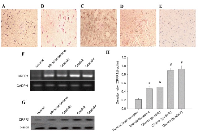
Figure 1.
CRFR1 mRNA and protein expression in glioma and normal brain tissues. (A-D) Immunohistochemical staining for CRFR1 in glioma specimens and (E) normal cerebral tissues. The CRFR1-positive cells contain homogeneous brown-yellow areas in the cytoplasm. (A) Medulloblastoma; (B) grade II; (C) grade III; (D) grade IV; and (E) normal cerebral tissue (magnification, ×200). (F) The CRFR1 mRNA levels in human glioma samples and normal cerebral tissues were detected using semi-quantitative reverse transcription-polymerase chain reaction. (G) CRFR1 protein levels were detected using western blot analysis. (H) Densitometry analysis of CRFR1 bands relative to β-actin. *P<0.01, #P<0.001, glioma tissue vs. normal brain tissue. CRFR1, corticotropin releasing factor receptor 1.

