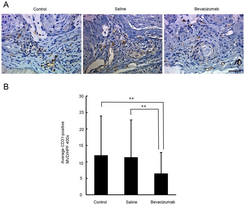
Figure 4.
Immunohistochemical assessment of CD31-positive microvessel density in xenograft tumors in response to bevacizumab treatment. (A) Representative microphotographs of CD31-immunostained tumor sections obtained from untreated control mice and from mice treated with bevacizumab or saline. Scale bar=50 µm. (B) Quantification of intratumoral microvessel density by CD31 immunostaining. Each bar represents the mean intratumoral microvessel density count ± SD. A significant decrease in microvessel density was observed in the tumors treated with bevacizumab compared with untreated control or saline-treated tumors (P<0.01). **P<0.01.

