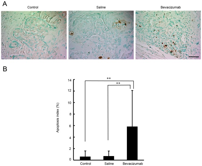
Figure 6.
Analysis of apoptosis indexes in tumor xenografts in response to bevacizumab treatment. (A) Representative microphotographs of TUNEL staining (brown). Sections were obtained from untreated control mice and from mice treated with bevacizumab or saline. Scale bar=50 µm. (B) Apoptosis index by TUNEL staining. Each bar represents the mean TUNEL-positive cell counts per total number of tumor cells ± SD. A significant increase in the percentage of apoptotic cells was observed with bevacizumab treatment compared with untreated control or saline-treated tumors (P<0.01). **P<0.01. TUNEL, terminal deoxynucleotidyl-transferase-mediated dUTP-biotin nick labeling.

