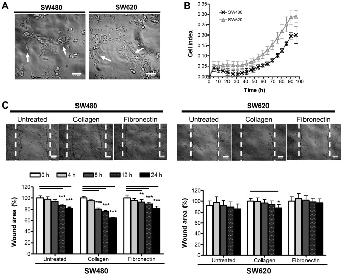
Figure 1.
Growth and migration characteristics of SW480 and SW620 colon cancer cell lines in vitro. (A) Adherent morphology of cells 24 h after seeding. Images were taken using a DSZ5000X inverted microscope and analysed using ImageJ software (scale bar, 0.1 mm). Arrows illustrate distinct cell morphologies. (B) SW480 and SW620 cell growth assessed over 96 h [biological triplicates using an xCELLigence™ real time cell analyzer (RTCA 1.2.1)]. CI represents a measurement calculated from the difference in impedance at an individual point of time (Zi) from the impedance at the beginning of the experiment (Z0), where CI = . (C) Migration potential of SW480 and SW620 cells over 24 h assessed by wound closure. Wound healing potential was measured according to the area of the wound (%) at 0, 4, 8, 12 and 24 h for SW480 cells and SW620 cells on untreated plastic, 1 µg/ml collagen type I or 5 µg/ml fibronectin. Dotted lines show the wound edges at 0 h. Results are representative of six independent biological replicates. Statistical significance in terms of differences in migration between the two cell lines was assessed in GraphPad Prism by two-way ANOVA with Bonferroni post hoc test. *P<0.05; **P<0.01; ***P<0.001. CI, cell index.

