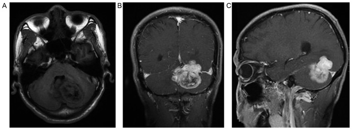
Figure 2.
Case 2. One of the features of a solitary fibrous tumor: The mass spanned the tentorium cerebelli. (A) Plain scan: The lesion signal was inhomogeneous; a low to intermediate mixed signal intensity was present on T1WI. (B) Enhancement scan: COR T1WI. (C) Enhancement scan: SAG T1WI. SAG, Sagittal plane; WI, weighted imaging.

