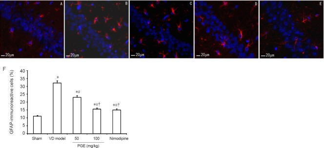
Figure 7.
Effect of PGE on hippocampal GFAP-immunoreactive cells.
(A–E) GFAP-immunoreactive cells were examined in the hippocampal CA3 region by immunofluorescence labeling. GFAP-positive cells are labeled red, and the labeling was mainly localized to the cytoplasm. Scale bars: 20 μm. (A) Sham group. (B) VD model group. (C) 50 mg/kg PGE group. (D) 100 mg/kg PGE group. (E) Nimodipine group. (F) Quantitation of GFAP-immunoreactive cells. Data are expressed as the mean ± SD (n = 10; one-way analysis of variance followed by Student's t-test). *P < 0.05, vs. sham group; #P < 0.05, vs. VD model group; †P < 0.05, vs. 50 mg/kg PGE group. PGE: Panax ginseng extract; VD: vascular dementia; GFAP: glial fibrillary acidic protein.

