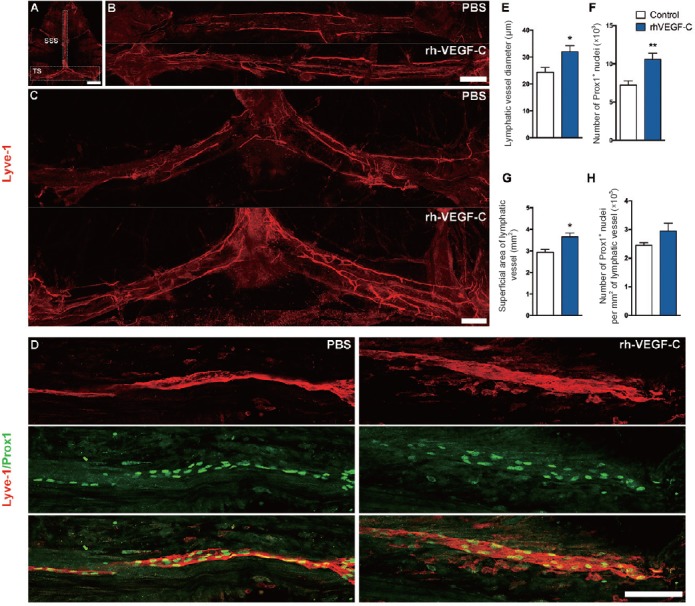
Figure 3.

Elevated dural lymphangiogenesis in transgenic mice injected with rhVEGF-C.
(A–H) Analysis of dura mater lymphatic vasculature in transgenic and experimental mice. (A) The dLVs along the SSS and TS, and the location of the images presented in (B) and (C). (D) Representative images of Lyve-1 and Prox1 labeling of meninges on day 7 after the last injection. Scale bars: 1000 μm (A), 500 μm (B, C) or 100 μm (D). (E–H) Quantification of the meningeal lymphatic vessel diameter (E), number of Prox-1+ nuclei located in dLVs (F), total superficial area of dLVs (G), and number of Prox-1+ nuclei per mm2 of lymphatic vessel. Data are presented as the mean ± SEM (n = 6; two-tailed Student's t-test). *P < 0.05, **P < 0.01, vs. control group. rhVEGF-C: Recombinant human vascular endothelial growth factor-C; SSS: superior sagittal sinus; TS: transverse sinus; dLVs: dural lymphatic vessels.
