Figure 3.

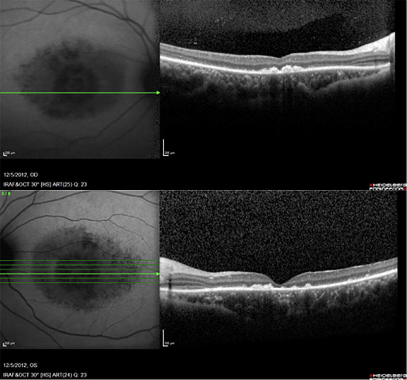
SD-OCT of both eyes at baseline shows foveal atrophy with disruption of the ellipsoid zone. Note that the ellipsoid zone is preserved nasally, demonstrating peripapillary retinal sparing.

SD-OCT of both eyes at baseline shows foveal atrophy with disruption of the ellipsoid zone. Note that the ellipsoid zone is preserved nasally, demonstrating peripapillary retinal sparing.