. 2015 Nov 2;7(1):128–135. doi: 10.1039/c5sc03053f

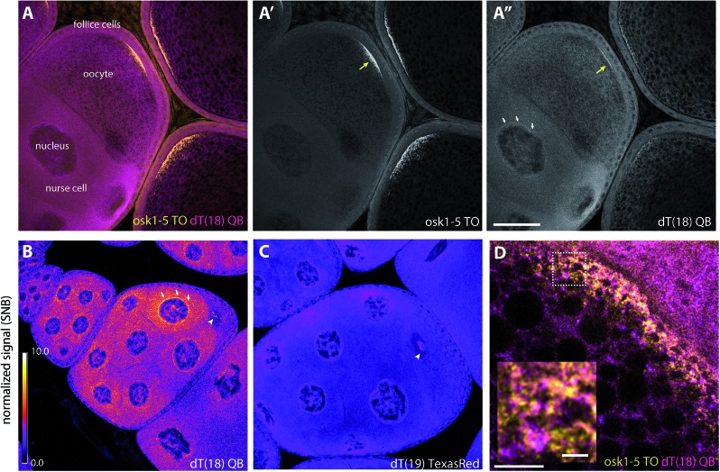
Fig. 6. Wash-free FISH of fixed Drosophila egg chambers. (A–A′′) Egg chambers comprising the translationally active nurse cells interconnected to the meiotic oocyte, surrounded by a layer of somatic follicular epithelium were co-labelled with a mixture of (A′) five TO-labelled oskar probes (osk-TO-LNA1-5, 0.1 μM each) and (A′′) dT(18) (0.5 μM) QB. (B and C) Normalized images of egg chambers labelled with (B) dT18-QB, (C) TexasRed-labelled dT19 (0.5 μM). Scale bars represent 50 microns (A–C). Normalization was done against the background signal measured within the oocyte nuclei (Fig. S10B, cyan dashed circle). White arrows indicate accumulated RNA around the nurse cell nuclei; yellow arrows indicate accumulation of oskar mRNA at the posterior pole; white arrowheads indicate speckles in the oocyte nucleus. (D) Super-resolution image (STED) of the oocyte posterior region of a stage 10 egg chamber, where oskar mRNA is enriched. The inset is an enlarged version of the image area within the dashed box. Scale bar represents five microns; scale bar in the inset represents one micron. Wash-free FISH of fixed Drosophila egg chambers.

Fig. 6