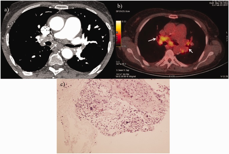
Fig. 3.
(a) Contrast-enhanced high-resolution CT of the chest showing lymph nodes and soft tissue in mediastinum (white arrows). (b) FDG-PET/CT axial slice at diagnosis showing active lymph nodes in mediastinum and lung hilum (white arrows). (c) Non-necrotizing granulomas consisting of epithelioid histiocytes, admixed with background of reactive lymphocytes and scattered plasma cells.

