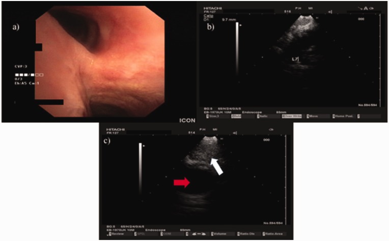
Fig. 4.
EBUS procedure. The patient was intubated with a STORZ rigid bronchoscope 12-mm outer rim and 11-mm inner rim diameter. The Pentax EB-1970UK endoscope was inserted through the working channel and multiple biopsies were taken from lymph node stations 7 (subcarinal) and 4 right with a 22-G EBUS needle. No endobrochial lesion was observed. (a) Camera of EB-1970UK EBUS showing the carina. (b) Ultrasound picture of EB-1970UK EBUS from EUB-6500HV Hitachi ultrasound source showing lymph node station 7 (subcarinal). (c) Ultrasound picture of EB-1970UK EBUS from EUB-6500HV Hitachi ultrasound source showing lymph node station 4 right, red arrow showing the superior vena cava and white arrow showing lymph node 4 right.

