Figure 1.
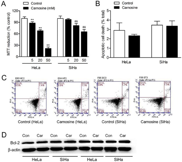
Effect of carnosine on cell viability in cultured HeLa and SiHa cells. Cells were treated with 5, 20, and 50 mM carnosine for 48 hours, respectively, and then the cell viability was assayed using the MTT reduction assay (A). Results were expressed as percentage of control, and were showed as mean ± SD. N = 10-12. **P < .01 versus control group in cultured HeLa cells, &&P < .01 versus control group in cultured SiHa cells. Apoptotic cell death was determined by PI and Annexin V-FITC staining followed by flow cytometry (B, C). Western blot analysis of the expression of Bcl-2 in HeLa and SiHa cells after carnosine treatment for 48 hours (D).

