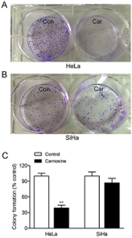
Figure 2.

Effect of carnosine on colony formation in cultured HeLa and SiHa cells. Cells were seeded at low density in DMEM supplement with or without carnosine (20 mM) for 14 days. The colonies were subsequently fixed with 4% paraformaldehyde and stained with crystal violet for analysis of colony formation. Representative images of the cloning wells, HeLa cells (A), SiHa cells (B). Quantitative image analysis of colonies in cultured HeLa and SiHa cells (C). Data were expressed as mean ± SD. N = 6. **P < .01 versus control group.
