Figure 1.
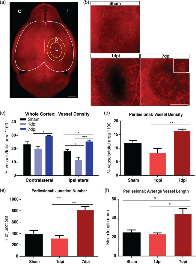
TBI results in vascular injury followed by repair. (a) Axial view of a vessel-painted brain from a Sham animal illustrating the dense vascular plexus. Analysis on the ipsilateral (I), contralateral (C) hemispheres, and perilesional tissue (P) around the lesion (L) was undertaken. Scale bar = 1000 µm. (b) Representative images from Sham, 1 dpi, and 7 dpi groups. Sham animals exhibited minor vascular disruptions due to the craniotomy. In the 1 dpi group, there was a clear disruption of the vessels that extended beyond the impact site. In contrast, the 7 dpi group showed the presence of newly formed vessels around the impact site. The inset illustrates these new vessels at high magnification. Scale bar = 1000 µm, 100 µm (insert). (c) Vascular analysis of the ipsilateral hemisphere demonstrating a reduction in vessel density (one-way ANOVA, p < 0.05) in 1 dpi group compared to shams, while the 7 dpi group showed a significant increase in vessel density (one-way ANOVA, ***p < 0.001, *p < 0.05) compared to the 1 dpi and sham groups. The contralateral hemisphere also exhibited a reduction in vessel density (not significant) at 1 dpi compared to the sham group. Interestingly, the 7 dpi group showed a significant increase in vessel density (one-way ANOVA, *p < 0.05) compared to the 1 dpi group with no change compared to shams. (d) Vascular analysis of perilesional tissue revealing a significant increase in vessel density (one-way ANOVA, **p < 0.01) in the 7 dpi compared to the 1 dpi group. (e) Analysis of the perilesional tissue revealing a significant increase in the number of junctions (one-way ANOVA, **p < 0.01) in the 7 dpi compared to 1 dpi and sham groups. (f) Similarly, we observed a significant increase in average vessel length (one-way ANOVA, *p < 0.05) in the 7 dpi compared to 1 dpi and sham groups.

