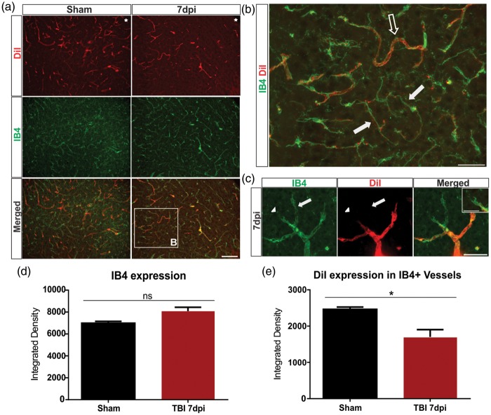
Figure 3.
Identification of perfused and newly formed blood vessels in the cortex. (a) Sham animals showed uniform vessel staining with DiI in coronal sections (see Figure 2), whereas the 7 dpi group exhibited a reduction in labeled vessels adjacent to the injury site (top panel). Similarly, in shams, there was a uniform staining of IB4+ vessels with an apparent reduction in the 7 dpi group. Interestingly, there appeared to be an increase in co-localized DiI and IB4+ vessels in the sham group, with little co-localization in the 7 dpi group. The boxed area is enlarged in (b). Star indicates region of impact. Scale bar = 100 µm. (b) A IB4+/DiI+ vessel from a 7 dpi mouse (open arrow) that is perfused (DiI+). Immature vessels, IB4+/DiI−, appear narrower and elongated in shape and are likely not fully formed nor perfused (solid arrow). Scale bar = 50 µm. (c) High magnification image of a newly forming vessel branching from an existing vessel. A new vessel is seen sprouting from a pre-existing vessel (arrow). IB4+ endothelial tip cells extend their filopodia into the cortex (arrowhead). Insert illustrates the filopodia of an endothelial tip cell. Scale bar = 25 µm, 15 µm (insert). (d) Analysis of the perilesional tissue revealing an increase in IB4 expression in the 7 dpi (Student t-test, p = 0.09) compared to the sham group. (e) Analysis revealing a significant decrease in DiI-expressing IB4 + vessels in the 7 dpi (Student t-test, *p < 0.05) when compared to the sham group.

