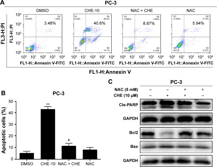
Figure 4.
CHE induced apoptosis via oxidative stress in prostate cancer PC-3 cells.
Notes: Cells were treated with CHE (10 μM) for 24 h in the presence or absence of prior 1 h incubation with NAC (10 mM). (A, B) Flow cytometry analysis of cell apoptosis using Annexin V-FITC+PI staining as described in the “Materials and methods” section. (B) Quantification of data presented in (A). (C) Western blot analysis for apoptosis-related markers in PC-3 cells. The statistic data for apoptosis cells were indicated and presented as mean ± SE from three independent experiments. **p<0.01 compared with the DMSO group. #p<0.05 compared with the CHE-10 group.
Abbreviations: CHE, chelerythrine; FITC, fluorescein isothiocyanate; PI, propidium iodide.

