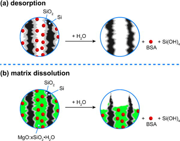
Figure 3.

Schematics depicting the two proposed mechanisms for protein release from (a) adsorption-loaded porous Si–SiO2 particles and (b) matrix-trapped porous Si–SiO2 particles. Images depict cross sections of the mesopores with bovine serum albumin (BSA) protein depicted with red spheres. (a) With adsorption-loaded porous Si– SiO2, the protein is weakly bound to the matrix and it is released prior to degradation/dissolution of the photoluminescent Si core. A burst release is observed, and photoluminescence from the Si skeleton degrades on a slower time scale. (b) With matrix-trapped porous Si– SiO2, the protein is embedded in a magnesium silicate matrix. Because the protein is trapped in this insoluble magnesium silicate phase, the release of protein is more closely tied to degradation of the carrier. A more controlled release is observed, and photoluminescence from the Si skeleton degrades on a similar time scale.
