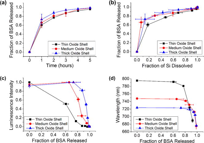
Figure 4.

Correlations between protein released, silicon dissolved, and photoluminescence from the Si skeletal core for adsorption-loaded core–shell porous Si–SiO2 particles as they undergo dissolution in aqueous base (0.1 M KOH). Experiments for three core–shell porous Si–SiO2 structures are displayed in each plot: Traces designated “thin oxide shell”, “medium oxide shell”, and “thick oxide shell” correspond to pSi particles oxidized at 700 °C for 25, 35, and 45 min, respectively. These samples were all “small-pore” microparticles, prepared to contain ∼10 nm pores (Table 1). (a) Fraction of bovine serum albumin (BSA) released as a function of time. (b) Fraction of BSA released as a function of silicon dissolved. (c) Integrated photoluminescence intensity (in range 650–800 nm) from particles as a function of BSA released. (d) Wavelength of maximum photoemission from the Si cores as a function of fraction of BSA released.
