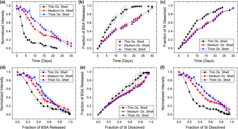
Figure 6.

Correlations between photoluminescence from the Si skeletal core, protein released, silicon dissolved, and time for trapping-loaded core– shell porous Si–SiO2 particles as they undergo dissolution in aqueous PBS (pH = 7.4) at 37 °C. The porous Si–SiO2 particles were prepared with a larger average pore size (∼20 nm), in contrast to the samples of Figure 5 (∼10 nm pores), which resulted in a substantially greater level of protein loaded (see Table 2). Protein trapping was achieved by means of precipitation of magnesium silicate as described in Figure 2, using 200 mM Mg2+. Traces designated “Thin Ox. Shell”, “Medium Ox. Shell”, and “Thick Ox. Shell” correspond to pSi particles where the skeletal core was oxidized at 700 °C for 15, 20, and 30 min, respectively, prior to protein/magnesium silicate loading. (a) Integrated photoluminescence intensity (in wavelength range 600–800 nm) from particles as a function of time. (b) Fraction of bovine serum albumin (BSA) released from the particles as a function of time. (c) Fraction of silicon dissolved as a function of time. (d) Integrated photoluminescence intensity from particles as a function of fraction of BSA released. (e) Fraction of BSA released as a function of silicon dissolved. (f) Integrated photoluminescence intensity as a function of fraction of silicon dissolved. Error bars represent standard deviation (n = 3).
