
Figure 4. Differential engagement of immune suppressive features in PDAC.
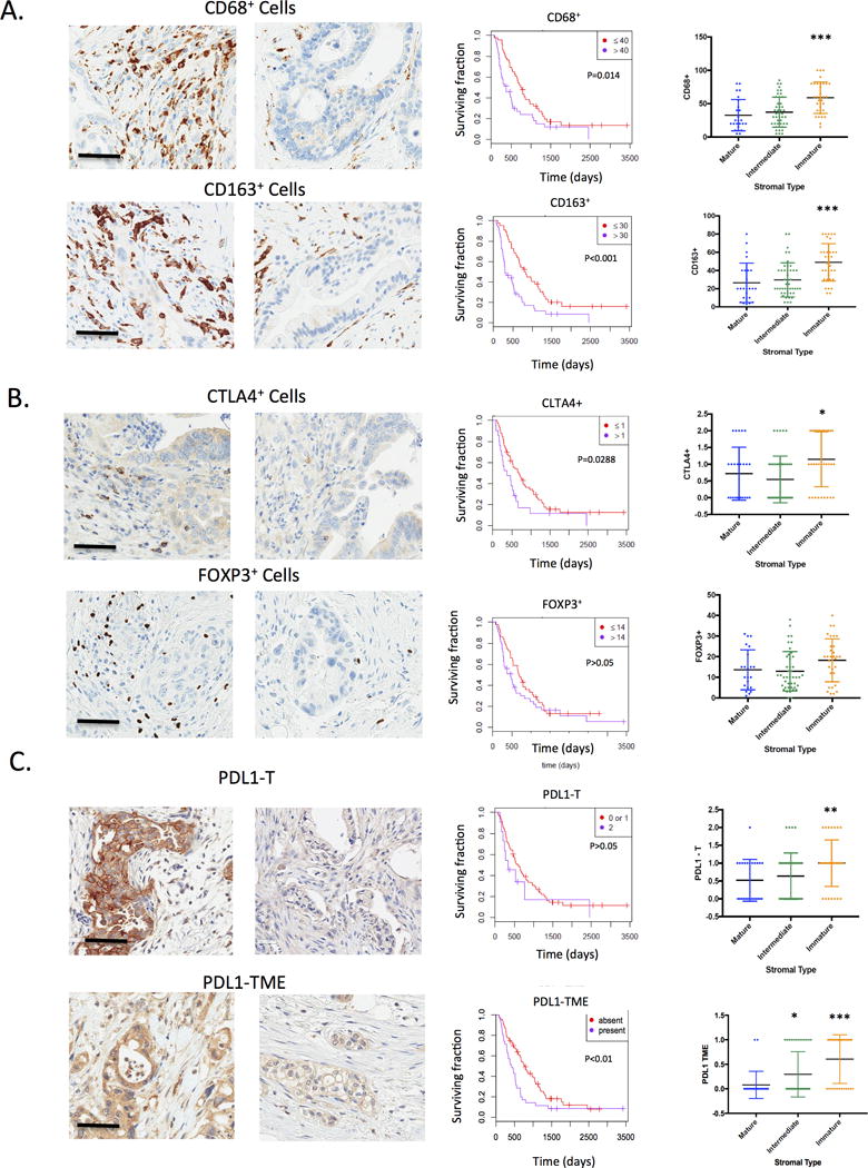
(A) The presence of macrophages or type II macrophages was determined by staining for CD68 and CD163 respectively. The overall presence of macrophages within the tumor microenvironment was significantly associated with overall survival as determined by Kaplan-Meier analysis. The level of CD68+ and CD163+ cells was associated with an immature stromal type (***p<0.001). (B) The presence of FOXP3 positive cells (indicative of T-regulatory cells) or CTLA4+ lymphocytes was determined within the PDAC tumors. CTLA4+ lymphocytes were significantly associated with overall survival, while FOXP3 was not significantly associated with overall survival as determined by Kaplan-Meier analysis. The level of CTLA4+ and FOXP3+ cells was determined as a function of stromal type (*p<0.05) (C) The expression of PDL1 was determined by immunostaining both in tumor cores (PDL1-T) and in the tumor micro-environment (PDL1-TME). The association with overall survival was determined by Kaplan-Meier Analysis. The level of PDL1 in various tumor comparments was analyzed as a function of stromal-type (*p<0.05,**p<0.01,***p<0.001).