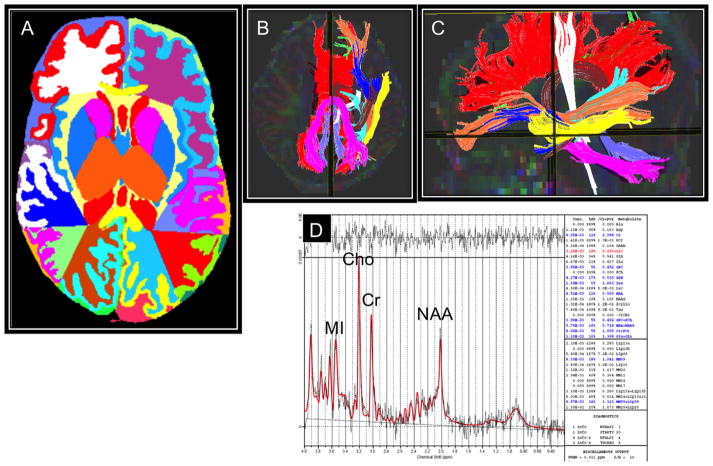
Fig. 1.
Examples of brain advanced MRI measurements including morphometry (A), diffusion tractography (B and C), and magnetic resonance spectroscopy (D). Representative advanced MRI examples at term-equivalent age display an extremely low-birth-weight infant’s brain that was segmented into tissues classes, subcortical structures, and lobes (A), 10 white matter tracts, displayed in axial and sagittal orientations (B and C), and a proton MRS spectrum displaying the four main metabolites, including N-acetylaspartate (NAA), creatine (Cr), choline (Cho), and myoinositol (MI).

