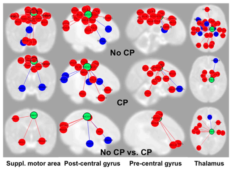
Fig. 3.

Functional connectivity MRI from 4 somatosensory and motor networks from 5 very preterm infants with cerebral palsy (CP) and 18 without CP. The columns and green circles represent the four sensorimotor regions of interest, including supplementary motor area, post-central gyrus, pre-central gyrus, and thalamus. The red and blue circles represent regions of the brain they are connected with; red signifies a positive correlation while blue represents a negative one. Infants with CP exhibited fewer sensorimotor connections (middle panel) than those without CP (top panel). The last panel displays several networks that were present in infants without CP (red connections) but were absent in infants with CP and a few hubs (blue) where infants with CP (blue) had more connections than those without CP.
