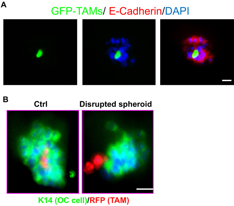
Figure 1. TAMs and OC cells in vitro 3D co-culture system were showed by Immunofluorescence.
A. TAMs and OC cells form spheroids in an in vitro 3D co-culture system. GFP+F4/80+CD206+ TAMs isolated from spheroids of ovarian cancer-bearing donor tomatolysM-cre mice and ID8 cells were co-cultured in the Matrigel-precoated 24-well plate for 48 h. The spheroids were subjected to immunofluorescent staining for E-cadherin for tumor cells. Images for GFP+ TAMs, E-Cadherin+ OC cells and DAPI for all cells in the spheroids are shown. B. Human TAMs were isolated and infected with lentivirus expressing RFP. RFP-expressing TAMs were incubated with SKOV3 human ovarian cancer cells followed by 3D co-culture for 72 h. Spheroids were immunostained with keratin-14. Scale bars = 10 μm.

