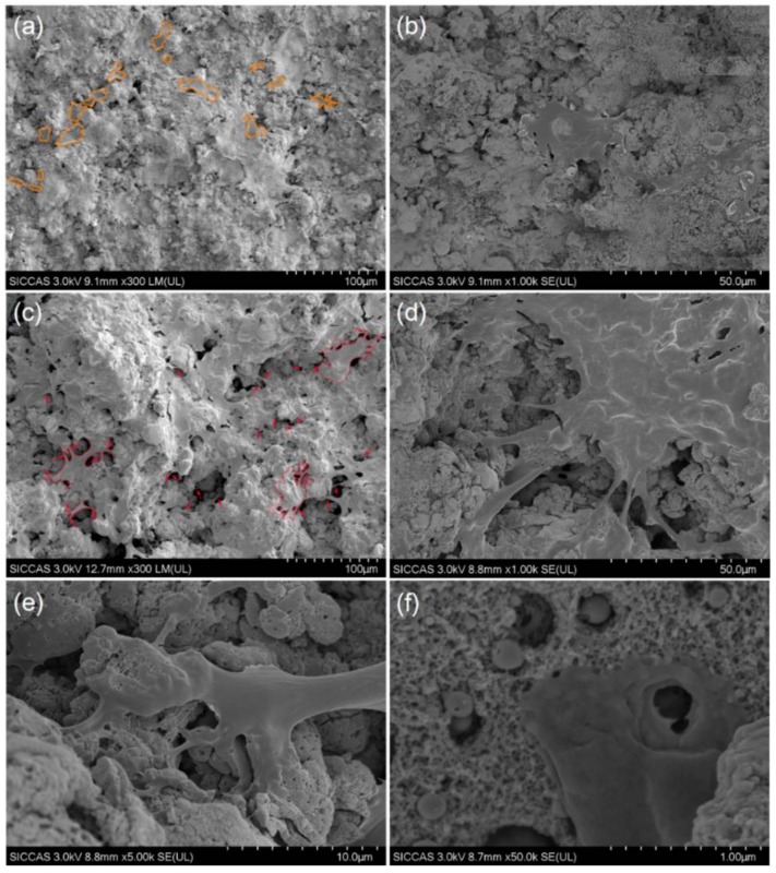
Figure 9.
SEM images of morphology of cells after 4 days on (a,b) MNT coating and (c–f) MNT coating under different magnifications. From (b), cell on MT coating presents spindle morphology with less stretched morphology. From (d), it shows fully spread cell with well-stretched morphology and a relatively large size on MNT coating. From (e), it presents a good interaction between the nanotube surface and the filopodia of hBMSC; From (f), a filopodia presents a good interaction with the MNT surface. (The orange outlines indicate the approximate morphology of cells on the surface of MT coating. The red arrows mark the filopodia of hBMSCs on MNT coating.).

