Figure 3.
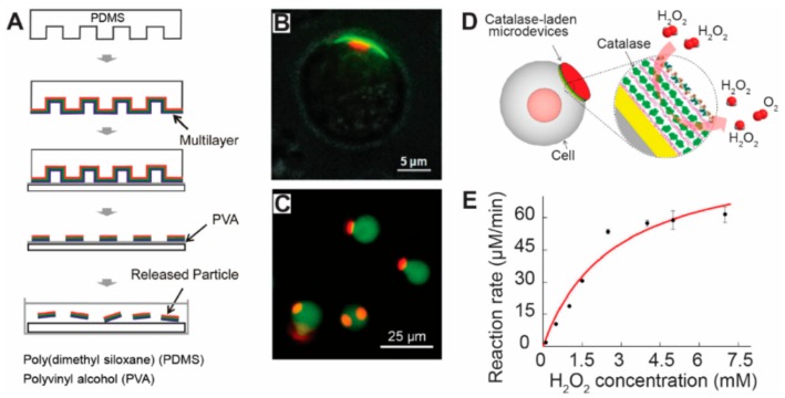
Soft lithography techniques were employed to generate micro/nanoparticles with controllable features for therapeutic delivery to cells. (A) Soft lithography techniques were used to generate disk-shaped micro/nanoparticles. Polyelectrolyte multilayers were assembled onto a soft PDMS polymer stamp. The stamps were then printed onto a substrate coated with PVA film, transferring the multilayer on the protruding pillars of the stamp onto the PVA film. Multilayer disk-shaped particles were produced by dissolving the PVA film with water [51], with copyright permission from © John Wiley and Sons; (B) Dot-on-pad micro/nanoparticles were attached onto a mammalian cell. The red dots were thermoplastic materials with dye. The green materials were the pad loaded with dye [125], with copyright permission from © John Wiley and Sons; (C) Disk-shaped particles were attached onto the cell surface. The red color indicates the particles with dye, and the green color represents the cells stained with another dye [128], with copyright permission from © American Chemical Society; (D) A schematic picture showing a cell-based drug delivery system. Catalase were laden into the disk-shaped particles. The catalase will react and transform H2O2 into O2 [128], with copyright permission from © American Chemical Society; (E) The reaction rate of the catalase depends on the concentration of H2O2 surrounding the cells in culture medium [129], with copyright permission from © American Chemical Society.

