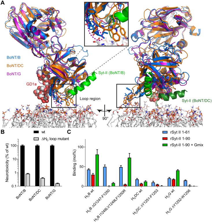
Fig 1. A hydrophobic loop is located between the synaptotagmin- and ganglioside-binding site of BoNT/B, DC, and G which confers high-affinity receptor binding and high toxicity of BoNT/B, DC, and G.
A Superposition of dual-receptor binding of BoNT/B (blue ribbon; PDB IDs 1EPW [55] and 4KBB [11]) to ganglioside GD1a (space fill model) and synaptotagmin-II (green ribbon), BoNT/G (pink ribbon PDB ID 2VXR [40]), and the interaction between BoNT/DC (magenta ribbon) and synaptotagmin-II (dark green ribbon side view; PDB ID 4ISR [22]) in proximity to a model membrane. Key amino acids of the hydrophobic loop (B: G1247-F1250, DC: Y1251-F1253; black box and insert) located between the ganglioside and synaptotagmin-binding sites are shown as stick models. B Determination of neurotoxicity of BoNT ΔHC loop vs wild-type in MPN assay. The neurotoxicity of single chain (sc) scBoNTBSL ΔG1247-F1250 (n = 4), scH6tBoNTDCS ΔY1251-F1253 (n = 4), and BoNTGS ΔY1252-W1256 (n = 2) was determined in comparison to the corresponding BoNT wild-type using dose-response curves (data are represented as mean ± SD). C GST pull down of wild-type and ΔHC loop-mutants of HCB, HCDC, and HCG by GST-synaptotagmin-II with or without gangliosides in Triton X-100 micelles. Binding of 100 pmol of the wild-type or ΔHC loop HC fragments to 150 pmol GST-rSyt-II 1–61, 1–90, or 1–90 in 20 mM Tris pH 8, 80 mM NaCl, 0.5% Triton X-100 in the presence of 125 μg of ganglioside mix (Gmix) embedded in Triton X-100 micelles immobilized to glutathione-sepharose 4B matrix and subsequent SDS-PAGE analysis (see also S2 Fig; data are represented as mean ± SD, n = 3).

