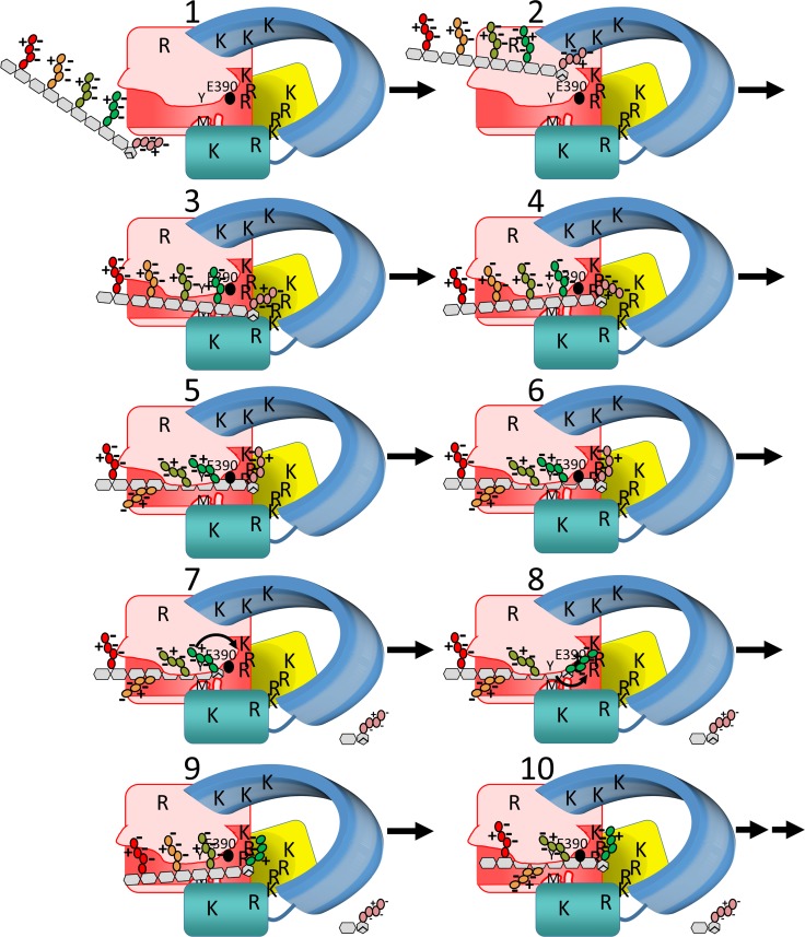
Fig 9. A proposed mechanism of PG hydrolysis by Cj0843.
The colors of the domains of Cj0843 are as in Fig 1. The 8 R/K residues in pocket 2 are indicated, and several additional R/K labels are drawn for illustrative purposes. In addition to E390 (black sphere), M410 and Y463 are labeled ‘M’ and ‘Y’, respectively. A narrowing of the active site groove is depicted in states 6–8 with an accompanying shift of the flanking NU-domain. The boat conformation of MurNAc-1 is drawn in state 6. The tetrapeptide sections of the 5 PG disaccharide units are colored as in Figures F and K in S9 Fig and S3 Video.

