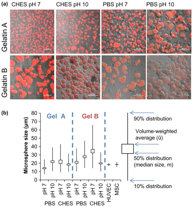
Figure 2.
Micrographs and size (b) measurements of crosslinked gelatin microsphere carriers. (a) Overlays of brightfield and fluorescent confocal microscopy sections of autofluorescent crosslinked gelatin microspheres. (b) Swelled microsphere dimensions in deionized water at room temperature. Box plots describe size distributions and median carrier diameter as a function of crosslinking regimen.

