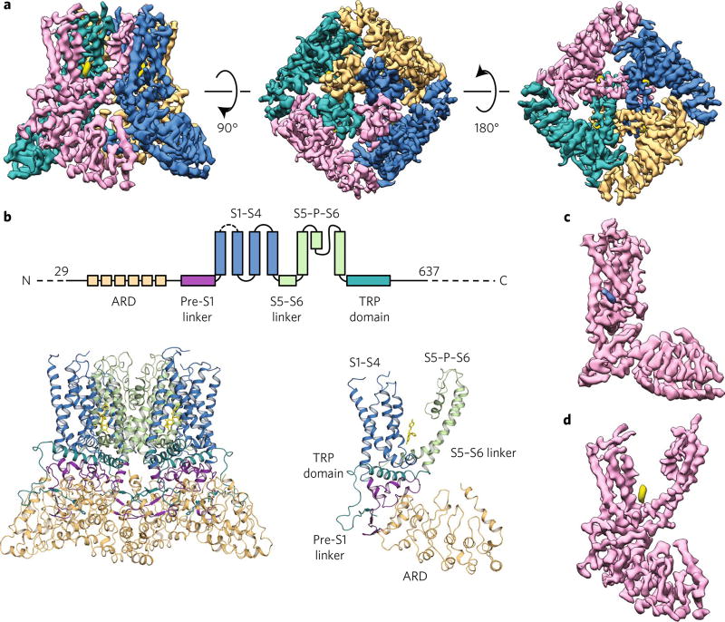
Fig. 1. Econazole-bound TRPV5 (TRPV5ECN) structure as determined by cryo-EM.
a, Density map of TRPV5ECN at 4.8-Å resolution. Each monomer is depicted in a different color (orange, blue, pink, and teal), and the density attributed to econazole is shown in yellow. The threshold of the density map was adjusted so that the putative calcium density is not visible for better visualization of the pore in this figure. b, A schematic representation of the domains present in TRPV5. Dashed lines indicate regions for which a model could not be built. Below, the TRPV5ECN model in tetrameric (left) and monomeric (right) form. c,d, A density map of a single monomer of TRPV5ECN (pink) with densities ascribed to phosphatidylinositol (blue) (c) and econazole (yellow) (d).

