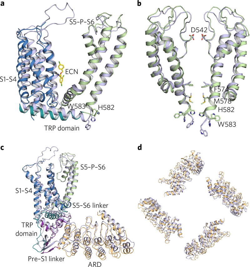
Fig. 5. Structural comparison of TRPV5ECN to TRPV6*.
a, Overlaid TM domains of TRPV5ECN (multicolored) and TRPV6* (blue). The regions of TRPV5ECN are labeled and colored based on the diagram in Fig. 1b. The histidine and tryptophan of interest in each pore are represented as sticks. The econazole molecule (ECN, yellow) from the TRPV5ECN structure is also presented as sticks. b, Aligned TRPV5ECN (green) and TRPV6* (blue) dimer pores. Residues of interest are labeled and shown as sticks. c, Aligned TRPV5ECN (multicolored) and TRPV6* (blue) monomers. The regions of TRPV5ECN are labeled and colored based on the diagram in Fig. 1b. d, Superimposed ARDs of TRPV5ECN (orange) and TRPV6* (blue).

