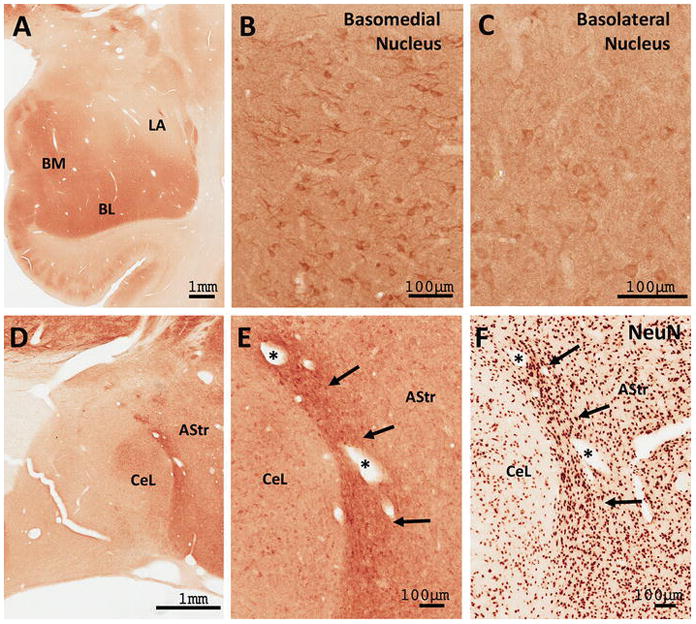
Figure 11. RGS14 is expressed throughout the monkey amygdala.

(A) Low power view of the distribution of RGS14 immunoreactivity in the monkey amygdala. (B–C) Light microscope images show discrete RGS14 staining of cell bodies and neuropil in the basomedial (BM) and basolateral (BL) nuclei, but not the lateral (LA) nuclei. (D–E) RGS14 labeling in the central lateral (CeL) and amygdalostriatal region (AStr). The dense band of labeling adjacent to the CeL corresponds to a sub-region of the AStr (arrows in E and F) that contains a larger neuronal density than neighboring AStr regions as revealed by NeuN immunostaining (F).
