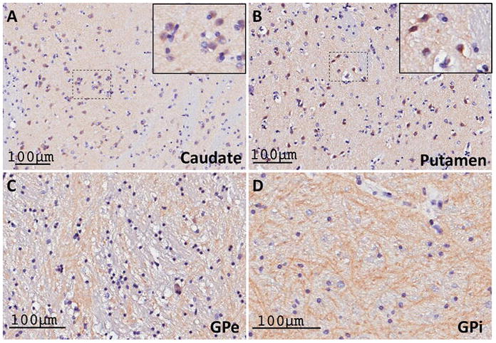
Figure 13. RGS14 expression in the human basal ganglia is consistent with labeling in the monkey basal ganglia.

(A–B) Light micrographs of RGS14-positive neuronal cell bodies, double stained with Nissl reagent, within a lightly labeled neuropil in the human caudate nucleus (A) and putamen (B). The insets in the upper right corner of each panel show examples of cell body labeling. (C–D) Dense RGS14-immunoreactive woolly fibers-like neuropil in the human GPe and GPi. The pattern of RGS14 labeling in both the striatum and the globus pallidus in humans is consistent with that described in monkeys.
