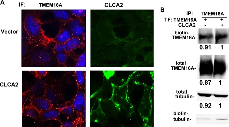
Fig 8. CLCA2 does not increase TMEM16A stability or surface occupancy.
(A) Immunofluorescence micrographs showing HEK293 cells stably expressing vector (upper row) or CLCA2 (lower row) and probed for transduced TMEM16A or CLCA2. Cells were fixed and stained with anti-Myc tag antibody for TMEM16A (red) or anti-FLAG antibody for CLCA2 (green). No difference in TMEM16A localization was observed. Scale bar, 20 microns. (B) Immunoblots from surface-biotinylated HEK293 cells stably expressing untagged CLCA2 or vector and transfected with TMEM16A-Flag. TMEM16A was immunoprecipitated, and the blot was probed with Alexa 680-tagged streptavidin. A second blot was probed for TMEM16A. Beta tubulin served as a control for protein concentration. To quantify expression, the fluorescence intensity was determined for each band and normalized to vector control using Licor software. To confirm that biotin labeling was confined to the surface, biotinylated proteins were precipitated with avidin-coated beads, blotted, and probed for beta tubulin, which produced only weak bands.

