. 2018 May 7;113(7):e180040. doi: 10.1590/0074-02760180040

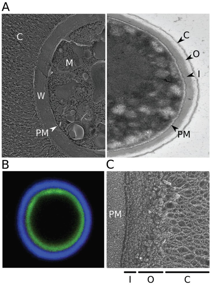
(A) Electron micrographs of Cryptococcus neoformans. Left, quick-freeze deep-etch image of cells grown in capsule-inducing conditions (as in Haynes et al. 2011); right, transmission image of cells grown in rich medium (YPD), which yields only thin capsules. C: capsule; W: cell wall; PM: plasma membrane; M: mitochondrion; O: outer cell wall layer; I: inner cell wall layer. (B) Fluorescent micrograph highlighting the cell wall and capsule. Cryptococcal cells were induced to form capsule and stained with florescein (green) to label the cell wall and monoclonal antibody 2H1 (blue, generously provided by Arturo Casadevall) to label the capsule, as in Pierini and Doering (2001). (C) Quick-freeze deep-etch image highlighting the two layers of the cell wall.

(A) Electron micrographs of Cryptococcus neoformans. Left,
quick-freeze deep-etch image of cells grown in capsule-inducing conditions (as
in Haynes et al. 2011); right,
transmission image of cells grown in rich medium (YPD), which yields only thin
capsules. C: capsule; W: cell wall; PM: plasma membrane; M: mitochondrion; O:
outer cell wall layer; I: inner cell wall layer. (B) Fluorescent micrograph
highlighting the cell wall and capsule. Cryptococcal cells were induced to form
capsule and stained with florescein (green) to label the cell wall and
monoclonal antibody 2H1 (blue, generously provided by Arturo Casadevall) to
label the capsule, as in Pierini and Doering
(2001). (C) Quick-freeze deep-etch image highlighting the two layers
of the cell wall.