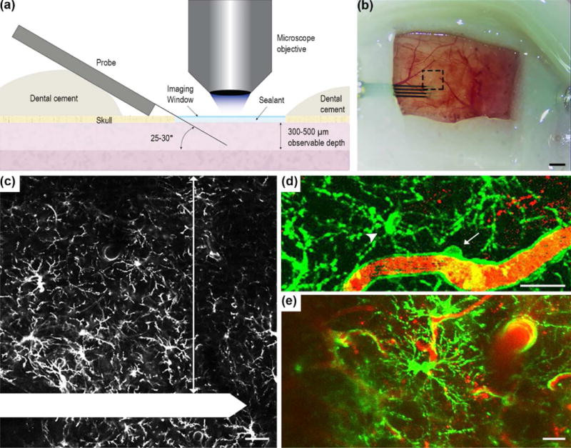
Figure 1. Experimental setup for in vivo tracking of NG2 glia and microglia cell dynamics.

(a) Schematic of chronic imaging window preparation for two-photon microscopy. The electrode was implanted at a 25-30° angle over the mouse visual cortex, sealed with kwik-sil and a cover glass was applied over the top to preserve imaging clarity and maintain tissue health. Dental cement was used to secure the probe in place and seal the craniotomy. (b) The chronic imaging window. Black dotted box is the region of interest where z-stacks were acquired. Scale bar = 1 mm. (c) Inset of (b). NG2 glia was observed and quantified 300 μm adjacent to the electrode shank (arrow). The electrode shank is outlined in white. Scale bar = 50 μm. Both vascular bound NG2+ pericytes (d, arrow), which display distinct morphology compared to NG2 glia (d, arrowhead), and proliferating NG2 glia (e) were excluded from quantification. GFP fluorescence is labeled green and blood vessels are labeled red for (d) and (e). Scale bar = 25 μm.
