
Figure 8.
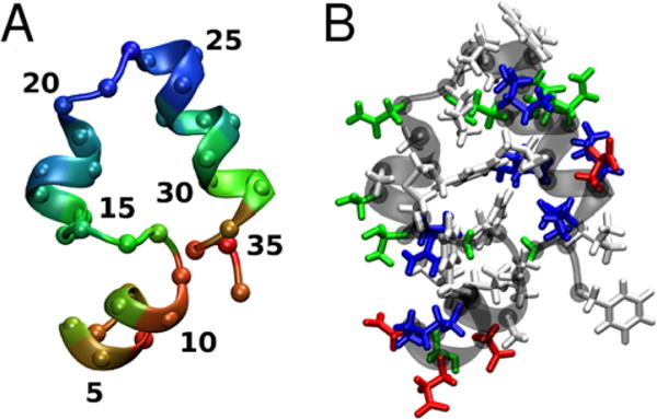
Structure of villin colored according to the average Cα distance to nearby villin molecules (A) (short: red, medium: green, long: blue) and by residue type (B) (hydrophobic: white, polar: green, acidic: red, basic: blue). Residue numbers 1–36 correspond to 41–76 in the biological sequence.