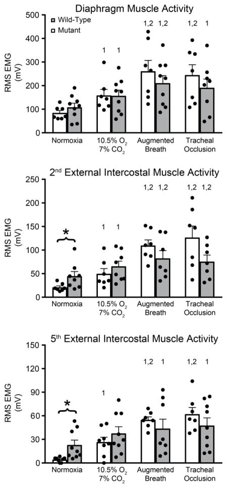
Figure 3.
Raw root-mean-squared EMG amplitudes (time constant: 50 ms): A. Diaphragm EMG amplitudes were comparable between WT and MT rats across conditions. B. 2nd external intercostal EMG amplitude more than doubled in SOD1G93A versus WT rats during normoxia (p<0.05) C. 5th external intercostal EMG amplitude quadrupled in SOD1G93A MT versus WT rats in normoxia (p<0.05). *p<0.05 Wild type vs. Mutant. 1p<0.05 compared to normoxia. 2p<0.05 compared to 10.5% O2-7% CO2.

