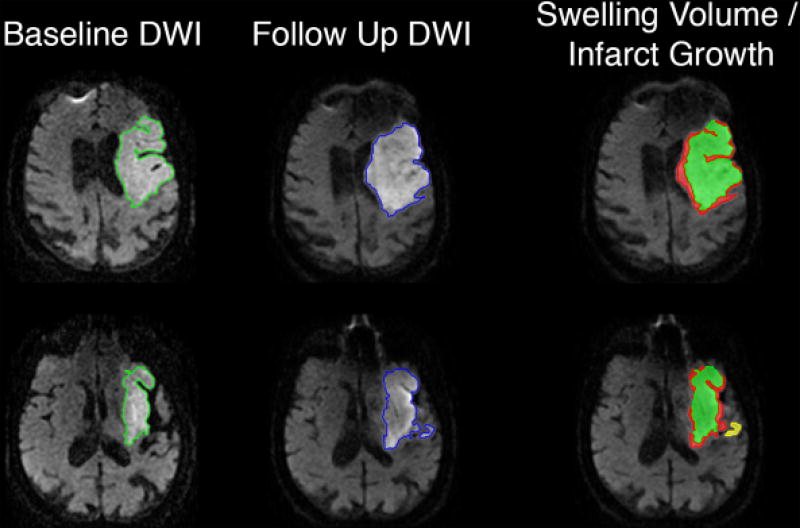
Figure 1.
Swelling volume was calculated using our previously described methods. Briefly, the baseline and follow up B0 sequences were co-registered using SPM, with the same transformation then applied to DWI sequences. The region of DWI hyperintensity was outlined on both baseline (green) and follow up (blue) MRIs, using a semi-automated seed-based technique. The follow up image was compared to the baseline in axial, coronal and sagittal planes, and any diffusion abnormality in a distinct anatomic region was defined as infarct growth (yellow). Finally, the baseline DWI lesion volume was subtracted from the resulting map of follow up DWI lesion and infarct growth, with any residual volume classified as swelling (red).

