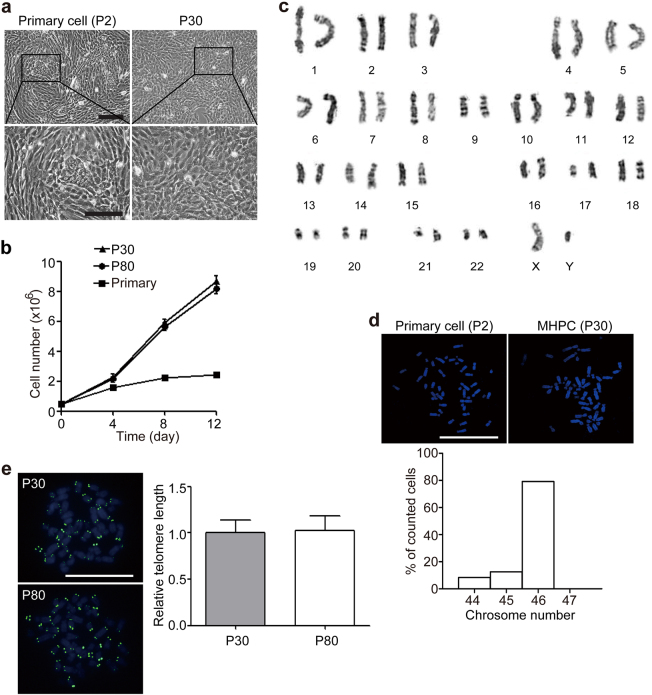
Fig. 1. Establishment of immortalized marmoset fetal liver cells.
a Morphology of immortalized marmoset fetal liver cell line MHPCs at passage 30 (scale bar = 100 µm). b Growth curve of MHPCs at passages 30 and 80, and isolated primary cells. Primary refers to isolated primary cells. Data represent mean ± sem (n = 3). The calculation was based on the online software http://www.doubling-time.com/compute.php. c G-band staining analysis of MHPCs at passage 30 showed a normal 46 XY karyotype. d Confocal microscopic images for DAPI staining of the choromosomes during mitosis. e Measurement of telomere lengths in MHPCs at passages 30 and 80 with Q-FISH. Data represent mean ± sem (n = 30)

