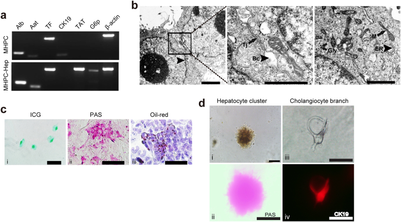
Fig. 3. In vitro evaluation for the bipotency of MHPCs.
a RT-PCR analysis of hepatocytic specific markers for differentiated MHPCs. MHPC-Hep refers to MHPC-derived hepatocytes. β-actin was used as a loading control. b Ultrastructure of MHPC-derived hepatocytes. Arrowheads indicate bile canaliculi (Bc) and endoplasmic reticulum (ER); arrows indicate tight junction (Tj), glycogen granules (Gly), and mitochondria (M) (scale bar = 1 µm). c In vitro functional evaluation of MHPC-derived hepatocytes, including (i) indocyanine green (ICG) uptake, (ii) PAS staining for glycogen storage, and (iii) Oil Red O staining for lipid accumulation. d In vitro bipotency of MHPCs, including (i) hepatic differentiation with 20 ng/ml OSM induction on 2D matrigel showed doughnut-like hepatocyte cluster morphology, and (ii) PAS staining for glycogen storage in MHPC-derived hepatocytes, (iii) branching structure of cholangiocytes formed by culturing in 3D type 1 collagen gel culture system, and (iv) CK19 staining (scale bar = 100 µm)

