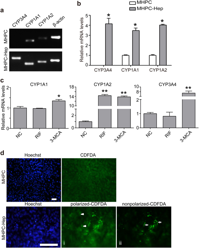
Fig. 4. MHPC-derived hepatocytes possess CYP enzyme activities and biliary secretion function.
a RT-PCR analysis for the levels of CYP enzyme expression in MHPC-derived hepatocytes. β-actin was used as a loading control. b Quantitative analysis of the mRNA levels of CYP genes by qPCR for MHPC-derived hepatocytes without inducer treatment. MHPC-Hep refers to MHPC-derived hepatocytes (n = 3, two-tailed t-test, *P < 0.05). c The mRNA levels of the CYP enzymes by qPCR, including CYP3A4, CYP1A1, and CYP1A2 in MHPC-derived hepatocytes after the induction with rifampicin (RIF) and 3-methylcholanthrene (3-MCA). Fold changes were normalized to the levels in the cells without induction, respectively (n = 3, two-tailed t-test, *P < 0.05; **P < 0.01). d CDFDA staining showed the accumulation of green CDF in functional bile canaliculi on the apical surface of MHPC-derived hepatocytes (scale bar = 100 µm). (i) Arrowheads point to CDFDA inside cells; (ii) arrows point to CDFDA around membrane

