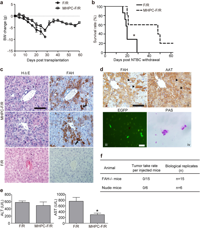
Fig. 5. Engraftment of MHPCs into Fah−/− (F/R) mice.
a Body weight change of transplanted F/R mice. Body weight was measured twice every week in F/R mice after transplantation of 1 × 10⁷ MHPCs. Body weights at the indicated time were normalized to the weights prior to the transplantation. BW body weight. b Kaplan–Meier survival curves of MHPC-transplanted F/R mice (n = 5) and F/R mice that did not receive cells (n = 7) after NTBC withdrawal (two-tailed t-test, *P < 0.05). c Immunostaining for FAH in liver tissues from transplanted F/R mice. The results indicated the integration of MHPCs in F/R livers and showed normal hepatocyte morphology in H&E serial sections. Arrows indicate binucleated hepatocytes (scale bar = 100 µm). d Immunostaining for AAT in liver tissues and in vitro functional evaluation of isolated MHPC-derived hepatocytes from transplanted F/R mice. (i, ii) Serial staining for FAH and AAT in liver tissues from transplanted F/R mice. Arrows indicate the co-localization of FAH and AAT (scale bar = 100 µm). (iii) Isolated EGFP-positive cells showing glycogen storage stained by PAS (iv) (scale bar = 100 µm). e Levels of serum ALT and AST in transplanted and control F/R mice (n = 3, two-tailed t-test, *P < 0.05). The blood was collected prior to the experimental endpoints. f The tumorigenicity assay for MHPCs in vivo

