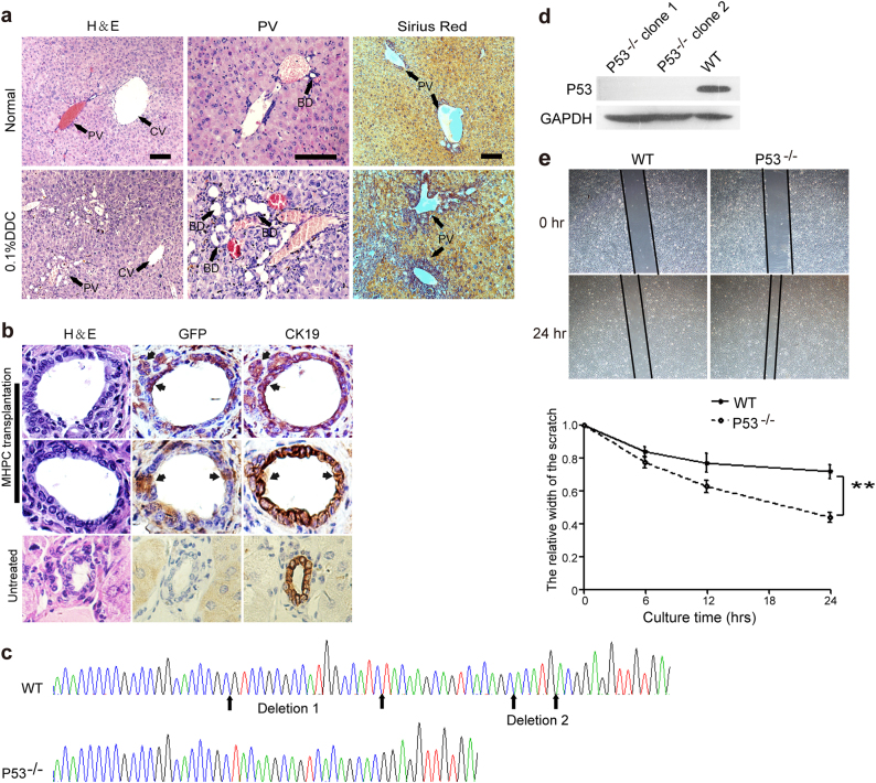
Fig. 6. Engraftment of MHPCs into the DDC-induced injury mice and genetic modification of MHPCs.
a H&E and Sirius Red staining of liver tissues from normal and 0.1% DDC-treated nude mice. Numerous small bile ducts (BD) appeared around the portal veins (PV) in the DDC liver (arrows). CV refers to central vein. b Serial immunostaining of GFP and CK19 in liver tissues from transplanted DDC-injured nude mice. The results showed the EGFP-positive MHPCs integrated in the large and small BD and expressed cholangiocyte marker CK19 (scale bar = 100 µm). c Sequence analysis of genetically modified MHPCs with CRISPR/Cas9 system for clone 1. Clones 1 and 2 showed identical deletions. WT refers to wild-type MHPCs. Two nonsense point mutations (A-C) and (C-G) were generated in the marmoset p53 gene. d Western blot analysis for the expression of p53 in transfected MHPCs. GAPDH was used as a loading control. A unit of 130 µg total protein was loaded. e Wound-healing assay for examining the migration rate of p53−/− MHPCs. A significantly faster migration rate was detected in p53−/− MHPCs than WT MHPCs (n = 5, two-tailed t-test, **P < 0.01)

