Figure 6.
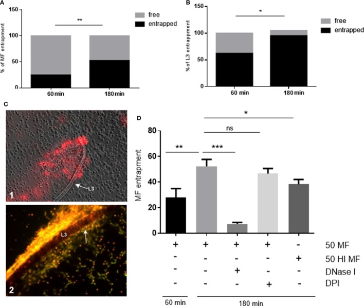
Dirofilaria immitis-induced parasite entrapment. Canine polymorphonuclear neutrophils (PMN) were exposed to vital D. immitis microfilariae (A) or L3 (B,C) for 60 and 180 min. (D) In parallel settings, the same number of PMN was incubated either with heat-inactivated (HI) microfilariae (HI MF) or pretreated with diphenyleneiodonium (DPI) before exposure to vital microfilariae. Furthermore, DNase I was added at the moment of exposure to vital microfilariae. Larvae were considered as entrapped when PMN and/or PMN-derived neutrophil extracellular traps (NETs) were in contact with larvae. The data are expressed as percentage of entrapped larvae relative to the total amount of larvae per condition. The formation of “clasp”-like NET structures sticking mainly to the anterior part of the larvae is displayed in white arrow: image [(C), 2].

