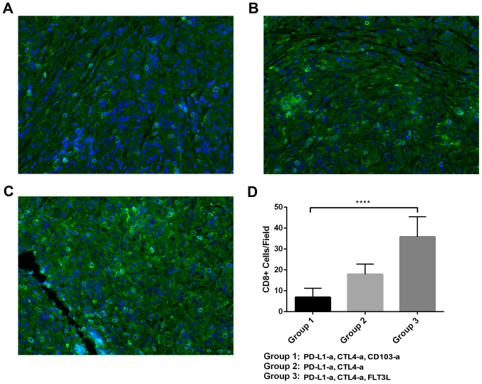
Figure 4.
Tumor infiltrating CD8+ T cells in a RCC xenograft mouse model. (A-C) Representative images of CD8+ T cells in the tumor tissue of RCC mice treated with ICBT and anti-CD103 antibodies, ICBT alone, or ICBT and FLT3L. Magnification, ×400. (D) Quantitative analysis of CD8+ T cells in the RCC xenograft mouse model. RCC, renal cell carcinoma; ICBT, immune checkpoint blockade therapy; -a, antibody; PD-L1, programmed cell death ligand 1; CTLA4, cytotoxic T-lymphocyte-associated protein 4; FLT3L, Fms-related tyrosine kinase 3 ligand; CD, cluster of differentiation. ****P<0.0001.

