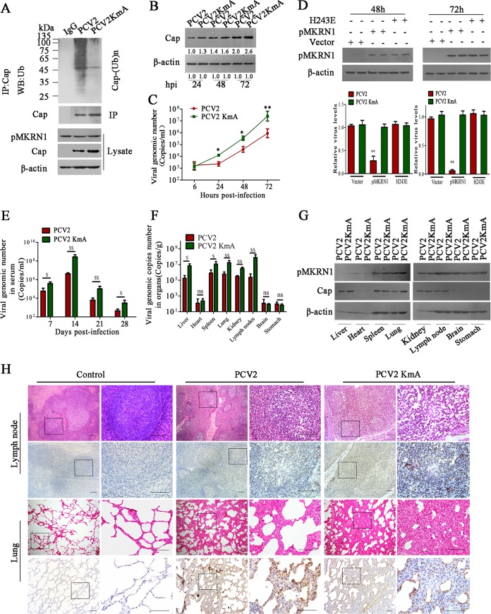
FIG 8.
Mutation of ubiquitination sites of Cap protein enhances the replication and pathogenesis of PCV2. (A to D) Mutant PCV2 (PCV2KmA) containing the three lysine residues (164, 179, and 191) replaced by alanine shows increased Cap levels and viral reproduction due to the loss of ubiquitination. PK-15 cells were infected with PCV2 or PCV2KmA, and Cap ubiquitination was examined (A), Cap expression levels were determined (B), and viral copy numbers were measured by quantitative PCR (C). *, P < 0.05 versus PCV2-infected cells; **, P < 0.01 versus PCV2-infected cells. (D) 64PKpmkrn1−/− cells were transfected with wild-type pMKRN1, the pMKRN1(H243E) mutant, or the pCI-neo vector, and then the cells were infected by PCV2 or the PCV2KmA mutant for 48 h and 72 h. Relative PCV2 or PCV2KmA levels were measured by quantitative PCR. **, P < 0.01 versus pCI-neo vector-transfected cells. (E to G) Piglets were infected with PCV2 or PCV2KmA (5 × 10⁵ TCID50) by intranasal injection. The numbers of PCV2 copies in serum at the indicated times (E) and at 28 days postinfection of tissues (F) were measured by quantitative PCR. The pMKRN1 expression levels in tissues at 28 days postinfection were measured by Western blotting (G). $, P < 0.05; $$, P < 0.01; ns, not significant. Results were confirmed in three independent experiments. (H) Hematoxylin and eosin staining of lung and lymph node tissues and immunohistochemical staining with anti-ORF1 antibody of lung and lymph node tissues derived from PCV2- or PCV2KmA-infected piglets at 28 days postinfection. The streptavidin-biotin-peroxidase complex method with counterstaining with hematoxylin was used. The regions with black rectangles were amplified, and the images are shown on the right. Bars, 100 μm.

