FIG 7.
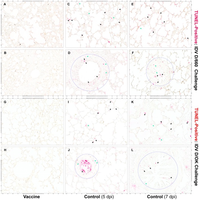
Micrographs of apoptotic cells in IDV-infected lung tissues detected with a TUNEL assay. TUNEL-positive epithelial cells lining alveoli (C, E, I, and K; red; black arrows) and bronchioles (D, F, H, and L; red; black arrows within blue circles) and nonepithelial cells (red, green arrows) in lung tissues were detected in all animals who received the sham vaccine (control) and subsequently challenged intranasally with IDV D/660 (C to F) or D/OK (I to L) but were not detected in animals who received the FluD-Vax vaccine and subsequently challenged with IDV D/660 (A and B) or IDV D/OK (G and H). Scale bars are shown on four sides of the image panels.

