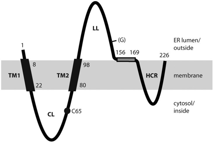
FIG 1.

Model for the transmembrane topology of the HBV S protein monomer in the ER membrane (shaded area). TM1 and TM2, transmembrane domains 1 and 2; CL, cytosolic loop; LL, luminal loop; HCR, hydrophobic C-terminal region; (G), facultative glycan N-linked to N146; C65, cysteine residue at position 65. Numbers refer to amino acid positions. The shaded rectangle represents a putative amphipathic helix. Domains in the ER lumen are located on the surfaces of secreted SVP, and domains in the cytosol are located inside SVP.
