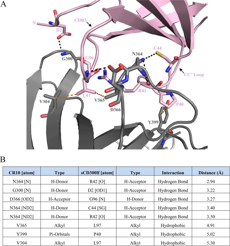
FIG 3.
Binding interactions of the murine norovirus CR10 P domain with sCD300lf. The CR10 P domain and sCD300lf are colored as in Fig. 2. (A) Close-up view of the binding interactions between the CR10 P domain and sCD300lf showing the direct hydrogen bond interactions (black lines) and hydrophobic interactions (orange lines). Residues on CDR3, the CC′ loop, and the N terminus of sCD300lf interact with the CR10 P domain. (B) List of residue interactions, where hydrogen bond distances were between 2.8 and 3.5 Å, while hydrophobic interactions were between 3.9 and 5.3 Å.

